

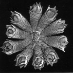
Living polyp of a Swiftia sp. sea fan. Photo credit: Peter Etnoyer, HRI.
Many deep-water animals have never been photographed alive in their natural habitat, they’re known only from their pickled state. Dried, dusty, and broken specimens fill museum drawers. “Living specimen photography” captures vital information before a specimen is collected. Remember to “snap” before you snip. It brings those dusty drawers to life!
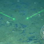
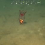
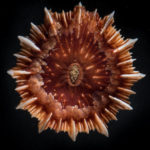
Swiftia sp. is a dark loving, azooxanthellate sea fan, one of many ‘asymbiotes’ in the twilight zone of the West Atlantic. The living colony’s color and morphology is seen below in a series of images through the microscope, and in-situ through the lens of the Sonsub Innovator ROV aboard NOAA’s RV Ron Brown on the 2003 Gulf of Mexico Deep Sea Habitats Expedition.


It gets EVEN better…there’s some microbial ecology data for this coral:
Bruck, T. B., W. M. Bruck, L. Z. Santiago-Vazquez, P. J. McCarthy, and R. Kerr, G. 2007. Diversity of the bacterial communities associated with the azooxanthellate deep water octocorals Leptogorgia minimata, Iciligorgia schrammi, and Swiftia exertia. Marine Biotechnology 9:561-576.
Note that both “Bruck”s have an umlaut over the “u” but the HTML kept interpreting it as a weird character so I took it out.
Awesome! I guess “zooming in” is not as hard as “zooming out” to show the landscape scale.
You can write
Mostly the shape and texture of some tiny microscopic bones inside the polyp called sclerites. This would be an order of magnitude finer scale than the polyp, and larger than the bacteria. The continuum is growing!
Thanks!